Research

Research Interest

Ultrasound Ultrafast Imaging

  • Ultrafast imaging (>6k fps)
  • High-speed motion tracking
  • Superresolution imaging
pic2

Shear Wave Elastography Imaging (SWEI)

  • Vessel stiffness imaging
  • Pulse wave velocity estimation
  • Reading of radiotherapy gel dosimetry
pic3

Microvasculature & Functional Ultrasound (fUS) Imaging

  • Ultrafast Doppler - High sensitive Doppler imaging
  • Functional US (fUS) imaging- Insights into Stroke and Neurovascular Diseases
  • Monitoring hemodynamics of brain diseases (stroke, PID, CSD...)
  • Tumor angiogenesis
pic4

US-guided High Intensity Focused Ultrasound (USgHIFU)

  • Monitoring by SWEI/Parameter imaging


Navigation system of Surgery

  • Ultrasound-guided surgery navigation system

Ultrasound AI

Ultrafast Imaging in Biomedical Ultrasound

The use of ultrasonic plane-wave transmissions rather than line-by-line focused beam scanning achieve ultrafast ultrasonic imaging with typical frame rate of higher than ~10k fps over a large field of view. In addition, the ultrafast images can be processed parallelly with graphical processing unit (GPU). This high speed imaging technique has inspired us to access dynamic tissue and flow, and other advanced imaging mode applications. 

Microvasculature Imaging (Functional Ultrasound)

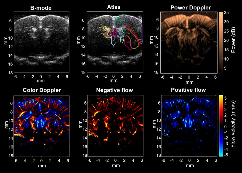
Ultrafast Doppler, also termed functional ultrasound (fUS), is an ultrasensitive and quantitative microvascular imaging technique that is able to access hemodynamics of target organs, expanding the field of application of ultrasound imaging and providing highly sensitive anatomical and functional mapping of vessels. With high frequency ultrasound, it can provide high spatiotemporal Doppler imaging (<100 μm, <1ms) to monitor perfusion change in small animals. We have developed a dynamic microvasculature imaging platform to study physiological functions of brain, liver, kidney, pancreas, and tumor etc. in the disease animal models. With this imaging tools, the perfusion responses under physiological stimulus, such as diseases pathogenesis, drug effects, … and so on can be investigated.

3D structure and microvasculature image of rat’s whole brain

Shear Wave Elastography Imaging (SWEI)

SWEI is a new ultrasound imaging tool capable to assess tissue stiffness. It uses an acoustic radiation pulse sequence to create shear waves, which propagate perpendicular to the ultrasound beam and are tracked with ultrafast imaging. Based on the mechanical model of elasticity-velocity relation, elastic modulus are calculated and reconstructed as elasticity 2D mapping. 

SWEI Demo for heterogeneous phantom


Shear wave propagation

Displacement map

Optical Coherence Elastography (OCE)

Shear wave elasticity strategy can also be applied on the optical coherence tomography (OCT) as non-contact elasticity imaging. Corneal biomechanics is critical for early diagnosis, optimal management of corneal diseases (e.g keratoconus) and for predicting the risks of surgical intervention of healthy corneas, such as post-LASIK ectasia. In this study, elasticity measurements of ex-vivo porcine corneal tissue are demonstrated with non-contact optical coherence elastography (OCE) by integrating ultraviolet (UV) laser pulse excitation and high speed phase-sensitive optical coherence tomography (PhS-OCT). The results respond linearly to intraocular pressure (IOP) (from 4-28 mmHg) based on the measurement of and group velocity (R2= 0.93) of propagating waves in the cornea launched by a single UV laser pulse. In addition, the 2-D elastic map of corneal tissue are obtained. This clinically-translatable OCE system can potentially generate personalized biomechanical models to help early diagnosis, and continued monitoring of keratoconus, pre-refractive screening for risks of ectasia, and treatment monitoring.